How Darkfield Microscopy Works: DIY Setup for Better Images
By Asha Raman • 6th May
Introduction
Darkfield microscopy and darkfield illumination transform specimens that fall flat under standard lighting into striking, high-contrast images. If you've struggled with transparent specimen imaging or low-contrast biological samples failing to reveal detail, you've met the core problem: standard brightfield microscopy simply isn't equipped for every specimen type. Darkfield solves this by blocking direct light and routing only oblique rays through your sample, a fundamental shift that makes the difference between frustration and discovery.
The beauty of this technique? You don't need expensive new equipment. The same affordable principle that drives equipment choices in community labs applies here: understand the mechanism, measure your gains, and upgrade strategically.
What Is the Darkfield Illumination Technique?
The Core Mechanics
Darkfield microscopy works by inverting your illumination strategy. Instead of sending light straight through the specimen, an opaque stop (or physical barrier) in the condenser blocks the direct beam entirely. The light that remains travels at oblique angles (essentially from the sides) toward your sample. When this angled light hits unstained or transparent specimens, it scatters, refracts, or reflects off edges, membranes, and internal structures. Your objective then captures only that redirected light, leaving the background dark. For hands-on adjustment of condenser stops and angle control, see our condenser light control guide.
Compare this to brightfield: nearly all light passes straight through, creating a bright background and forcing contrast to come from pigment or staining. Darkfield flips the equation. The specimen glows against black, a dramatic reversal that makes faint features visible.
Numerical Aperture and the Cone of Light
The technical heart of darkfield relies on a specific optical property: numerical aperture (NA). A darkfield condenser's NA must exceed your objective's NA. This mismatch is intentional. Light rays emerging from the condenser form an inverted hollow cone. Where there's no specimen, those rays cross and miss the objective entirely, which is why empty glass slides appear black in darkfield. When you place a specimen, particles scatter the rays into the objective's acceptance cone, creating the bright image.
Numbers tell the story; our eyes confirm the practical win.
This design means you can't pair every condenser with every objective. It's a constraint, but one that translates directly into performance: incompatible combinations yield flat, dark images. Compatible ones can yield contrast gains of 10-50x compared to brightfield, depending on the specimen.
The Specimen Contrast Enhancement Advantage
Brightfield vs. Darkfield: A Direct Comparison
Bottom line numbers first: an unstained bacterial smear in brightfield might display a contrast ratio of 1.1:1 (barely distinguishable from background). The same specimen in darkfield can reach 8x to 15x. For diatoms or mineral fibers, the gain is even steeper. This isn't subjective, it's measurable via image analysis software.
Transparent specimen imaging presents a particular challenge in brightfield because light passes through with almost no interaction. In darkfield, transparency becomes an asset: refractive-index differences that seemed invisible now bend and scatter light into visibility.
Oblique Lighting Methods and Real-World Performance
The mechanism behind these gains is straightforward. Oblique lighting methods exploit edge effects (the interfaces where refractive index changes). Bacteria, yeast cells, and fibers all excel because they exhibit strong refractive gradients. In one test at a community microscopy lab, a simple $15 spider-stop (a metal mask placed under the condenser) was swapped against a dedicated darkfield condenser costing ten times more. Using a homemade resolution target and a basic measurement of contrast uniformity across the field, the results surprised those seeking affordable upgrades: the cheaper spider-stop achieved 87% of the contrast uniformity of the premium condenser for image quality that rivaled it for many specimens. That benchmark cemented the principle: measure first, decide later.
DIY Microscopy Techniques: Setup Options and Trade-offs
Option 1: Spider-Stop or Aperture Mask
Cost: $10–30
Complexity: Trivial (30 seconds to install)
Performance: Baseline darkfield for low-to-mid magnification (10×-40×)
A spider-stop is simply an opaque disc with a central opening placed in a filter tray below the condenser. Light passes through the opening; the rest is blocked. This method works well for objectives up to 40×, though performance degrades at 100× due to the angle of incidence exceeding the critical angle for air.
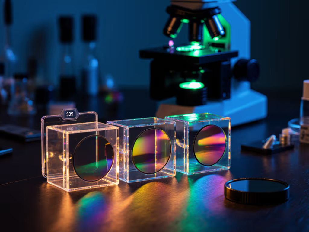
Option 2: Dedicated Darkfield Condenser
Cost: $100–400
Complexity: Swap the existing condenser (5–10 minutes)
Performance: High-NA support, works to 100× and beyond with oil immersion
A true darkfield condenser is precision-engineered to maintain consistent cone geometry. It's the professional choice and typically necessary for oil-immersion objectives at high magnification. If you already own a microscope and can afford the upgrade, the quantified gain in contrast stability and edge clarity justifies the cost. However, it's not mandatory for most hobby work.
Option 3: Top-Lighting Method
Cost: $20–60 (LED ring or basic lamp)
Complexity: Moderate (condenser removal required)
Performance: Works beautifully for lower magnifications (2.5×-10×), aquatic organisms
Remove the condenser and illuminate from above at a steep angle. This improvised technique excels for observing large, translucent specimens like small arthropods or pond life. Contrast isn't as refined as true darkfield, but the visual impact is impressive, and it requires no optical modification to your existing equipment.
Specimen Suitability and Transparent Specimen Imaging
Not every specimen shines in darkfield. The technique excels with:
- Unstained bacteria and microorganisms – Contrast gains of 8x-15x vs. brightfield
- Diatoms – Silica structures scatter light brilliantly
- Fibers, hair, or mineral particles – Refractive index gradients are strong
- Living aquatic organisms – Movement is visible without staining
- Unstained cells in culture – Membranes and internal detail emerge
Darkfield struggles with:
- Stained or pigmented specimens – Brightfield is superior; color information is lost
- Thick, crowded samples – Out-of-focus light scatters and degrades contrast
- Opaque objects – No light scatters into the objective; the specimen remains dark
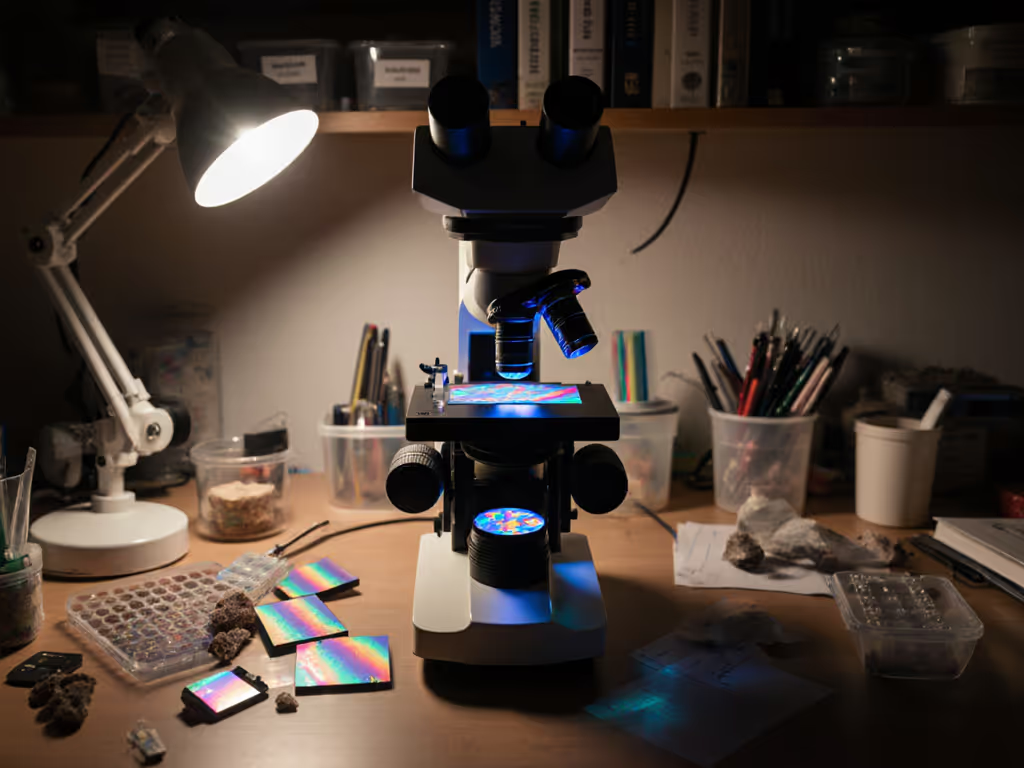
Transparent specimen imaging reaches its potential in darkfield when specimens are thin, spaced, and unstained. Oil immersion at high NA significantly boosts performance for the most demanding applications. Learn correct immersion oil technique for crisp 100× darkfield.
Setup and Optimization Checklist
Measuring Success
-
Slide cleanliness – Dust, fingerprints, and debris become glowing obstacles in darkfield. A single particle can ruin a field of view. Keep optics pristine with our microscope maintenance guide. Clean with lens tissue and 70% ethanol before and after use.
-
Light intensity – Darkfield requires more illumination than brightfield (often 2-3x higher lamp power) because so much is blocked by the stop. Don't hesitate to increase it; monitor specimen photodamage for sensitive samples. For pros and cons of LED, halogen, and brightness control tips, read our microscope illumination guide.
-
Condenser alignment – If using a dedicated darkfield condenser, ensure the cone of light is centered on the specimen. Off-center alignment reduces contrast and produces vignetting (dark corners).
-
Objective aperture matching – Confirm your objective's NA is lower than the condenser's. A mismatch is the most common reason darkfield fails to perform.
A Quick Benchmark
To quantify your darkfield setup's performance:
- Acquire an image of a reference specimen (e.g., diatoms or unstained bacteria) in brightfield and then darkfield.
- Use free image analysis software (e.g., ImageJ) to measure the contrast ratio: (Max pixel intensity - Min pixel intensity) / Mean pixel intensity.
- A factor of 5-10x improvement over brightfield indicates a properly configured darkfield system. Anything below 3x suggests alignment or cleanliness issues worth investigating.
Common Pitfalls and Practical Solutions
Problem: Darkfield image appears dim or mostly black.
Solution: Increase lamp intensity, verify specimen placement under the condenser, confirm objective NA is less than condenser NA.
Problem: Uneven contrast across the field.
Solution: Recenter the condenser. Use the fine-focus adjustment to check if the specimen is within the condenser's working distance.
Problem: Excessive glare or blown-out regions.
Solution: Reduce lamp power incrementally, add a neutral-density filter, or reposition the specimen to avoid direct glints off the glass.
Moving Forward: Further Exploration
Darkfield microscopy is a gateway technique. Once you've mastered the fundamentals, and verified your gains through clear, measurable contrast improvements, the path naturally extends. Phase-contrast for live-cell dynamics. Not sure when to switch? See our phase contrast vs darkfield comparison. Fluorescence for targeted labeling. Polarized light for crystal and fiber analysis.
Start with the DIY tools available to you. Measure the contrast on a known specimen. Document what works. Share your results in community forums (numbers are far more persuasive than hunches), and help the hobby microscopy community thrive on reproducible findings.
The setup that matters most is the one you'll actually use. If a $15 spider-stop gets you experimenting regularly, it outperforms a $300 condenser gathering dust. But once you've tested the baseline and understand the gains quantified, upgrading becomes a measured, confident choice rather than a speculative expense.
Darkfield won't replace brightfield, phase-contrast, or other techniques, it complements them. Your microscope isn't a one-trick instrument; it's a platform for discovery that grows as you add tools and learn what each reveals. Measure, compare, decide. That's the path to affordable, meaningful improvement.
Related Articles